Figure 2 shows the tube well positioned in the esophagus. On-screen visualization provides immediate feedback on tube placement which can reduce or eliminate the need for x-ray confirmation.
Cureus Hemothorax Following Traumatic Dobhoff Tube Insertion
Auscultation was performed in all 78.
. Confirmation of feeding tube placement. Open Access Case Report DOI. Confirmation of NGT or OGT placement by ultrasound was obtained in 23 patients.
Place tube through nares and ask patient to swallow as you pass the tube Giving the patient a cup of water with a straw may help IMPORTANT. A review of the x-ray showed that the feeding tube was in the main bronchus. Most tube positions are checked by assessing pH of tube aspirate normal tube descends the thorax in the midline tube bisects the carina tube crosses the diaphragm in the midline the tip sits below the diaphragm viewing the tube you need to be confident that you can see the tip most tubes are visible on a chest x-ray without a guide wire.
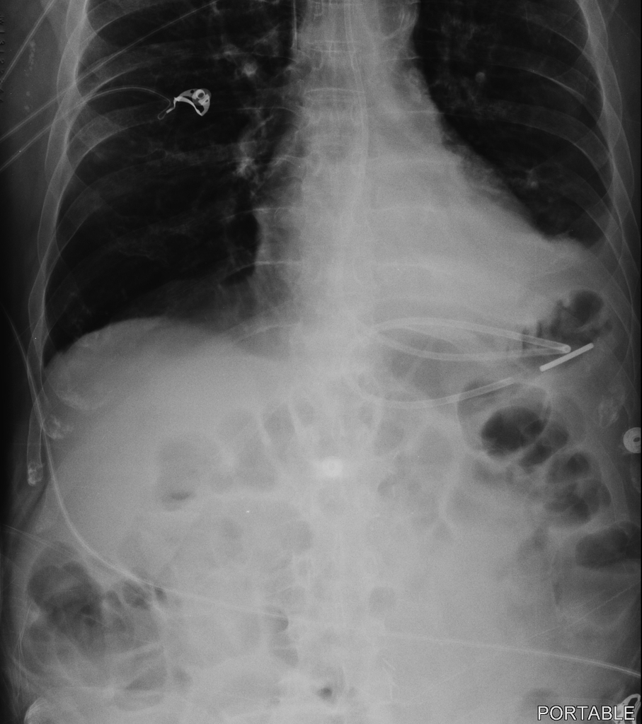
The distal tip of the feeding tube is in a loop of jejunum in patient status post gastrojejunostomy. Auscultation via air bolus aspiration of gastric contents and pH testing with litmus paper were not utilized to reconfirm placement since all of these methods have been proven to be unreliable PA-PSRS 2006. Abdominal X-ray after Dobhoff tube DHT placement to confirm accurate positioning.
Dobhoff tube is a special type of nasogastric tube NGT which is a small-bore and flexible so it is more comfortable for the patient than the usual NGT. The nurse is sure of gastric placement. Steps for NG Feeding Tube Placement in an Awake Patient Step 1.
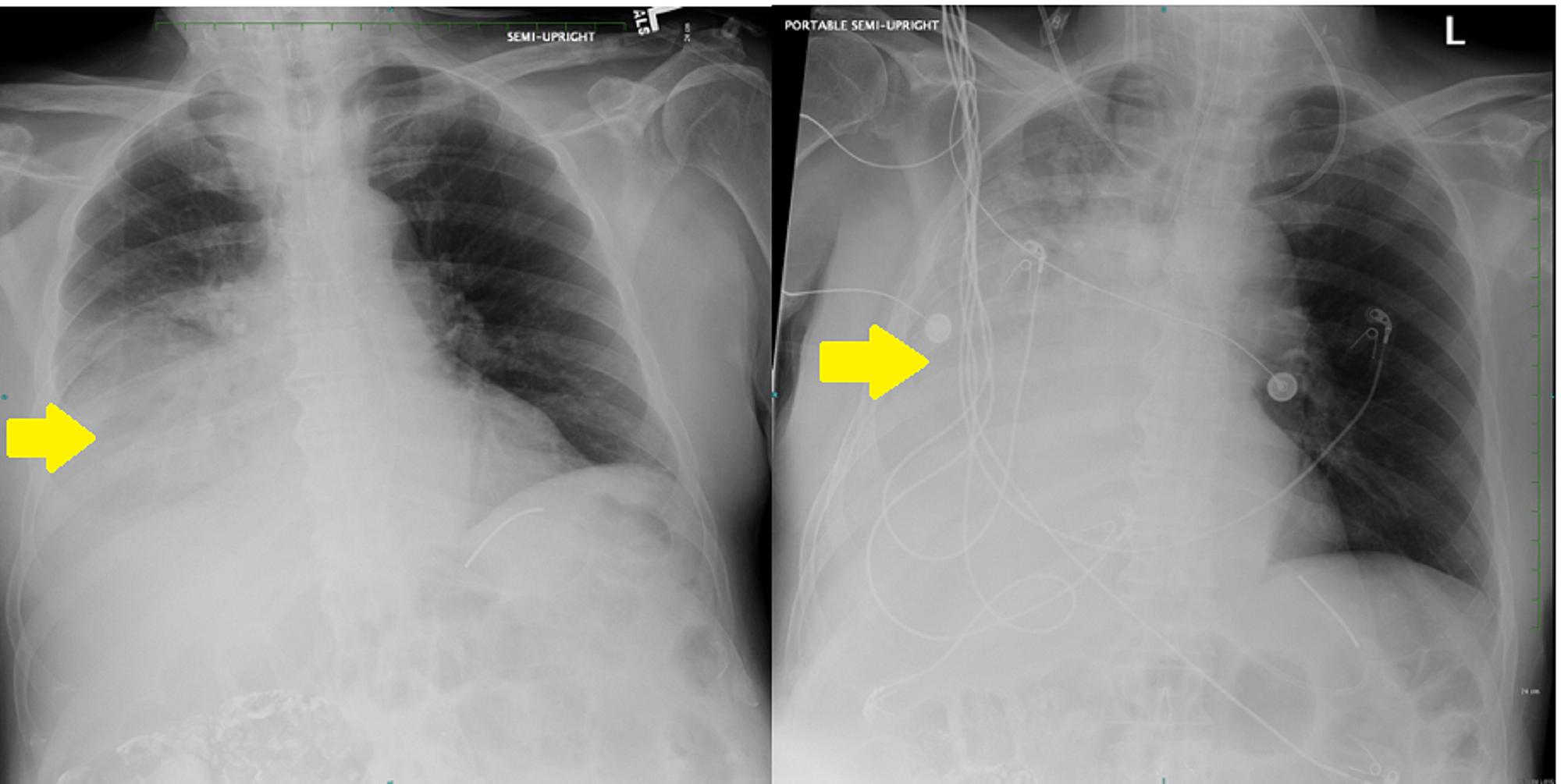
Tube feedings were initi-ated. If we had continued to advance the tube we would have risked causing a pneumothorax. Dobhoff tube is a special type of nasogastric tube NGT which is a small-bore and flexible so it is more comfortable for the patient than the usual NGT.
The x-ray was read and placement confirmed. Insufflate the stomach with 300 cc of air. A Bentson wire was used to.
Chest X-ray for confirmation of Gastric feeding tube placement 3148 Remove stylet-once placement has been confirmed by the physician and practitioner order is written stating that tube placement verified by xray and may be used. Measure tube from tip of nose to subxyphoidprocess about 3035cm in most patients Step 2. Tube to 30 centimeters and took a chest x-ray.
Confirmation that the feeding tube is properly placed in the stomach or small bowel involves documenting the following on a chest x. Of 78 nasoenteral intubations in 46 patients using a Dobbhoff Biosearch Medical Products weighted enteral feeding tube gastric aspirates were evaluated in 28. It is recommended to flush the tube with the stylet with sterile salineor water and gently remove.
Measure the small bore feeding tube SBFT from the nose tip down to the stomach and across the midline several centimeters approximating the location of the pylorus. What can go wrong. The tube is inserted by the use of a guide wire called the stylet see image1 which removed after the tube correct placement is confirmed.
Chest X-ray performed shortly after the tube placement demonstrated that the tip of the Dobhoff tube was within the right lung base following the course of the right mainstem bronchus Figure 1. All 26 patients had x-ray confirmation of NGT and OGTs correctly placed in the stomach. We told the nurse to check by air bolus while we await a second xray to be completed and read.
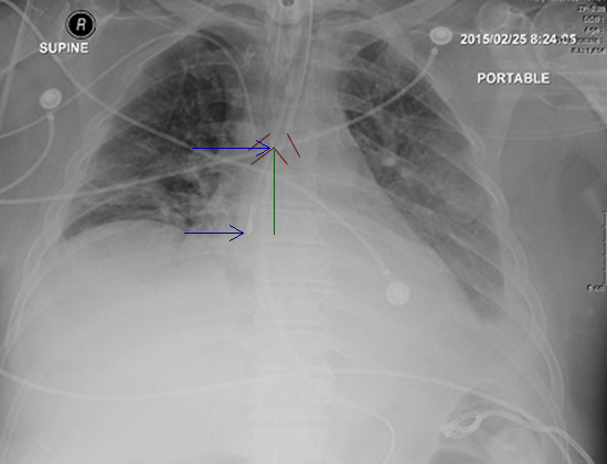
The wire was left in. As seen in Figure 1 the tip of the Dobhoff tube is in the left mainstem bronchus. It was a 12 french by the way.
ZEndotracheal placement zEpistaxis zSinusitis zEsophageal perforation zAspiration zPneumothorax. Dobhoff tubes are inserted into the stomach or the duodenum by way of nasal passage with the use of a guidewire called a stylet which is removed after confirmation of correct placement. Reconfirming Dohoff NG tube placement was completed by evaluating the daily chest x-ray or assessing the tube centimeter length.
The tube is inserted by the use of a guide wire called the stylet see image1 which removed after the tube correct placement is confirmed. Data was collected at initial placement prior to x-ray confirmation. Tube placement Ideally patient.
An x-ray can ensure that the Dobhoff tube has been placed correctly. The patient experi- enced respiratory dis-tress. 120cm preferred over blue tip dobhoff tube Lubricant 60 ml syringe.
Gastrointestinal anatomy before and after Roux-en-Y. In order to prepare a patient for the insertion of a Dobhoff tube the esophagus and nasal cavity are numbed and the patient if conscious may be given a mild sedative. An electromagnetic stylet provides real-time location information on the tube tip placement within a patients anatomy.
Dobhoff tubes have a metal weighted end composed of lead and wrapped in silicone that helps guide it through the gastrointestinal tract. Which tube is the most common NG tube. Microsoft PowerPoint - Nasogastric_tubes Author.
CORTRAK can be used to confirm bedside tube placement without x-ray. The tube is inserted by the use of a guide wire called the stylet see image1 which removed after the tube correct placement is confirmed. The Dobhoff tube was advanced through the right nasal cavity and the tube was confirmed to be in the distal esophagus.
We had a situation where a dubhoff did not show up anywhere on the x-ray. With CORTRAK 2 you can. A Dobhoff tube was placed by a house physi-cian.
Patients are usually positioned on the right side while the tube is put into the nose. If there isnt confirm placement with xray KUB zWith dobhoff tubes should always confirm placement as no suction will be applied. For these 23 patients POCUS agreed with radiographic findings.
For 3 patients the NGT and OGT was unable to be identified by ultrasound. The tube was removed and re-advanced. At our facility we x-ray all feeding tubes for placement verification.
Dobhoff tube is a special type of nasogastric tube NGT which is a small-bore and flexible so it is more comfortable for the patient than the usual NGT. Dobhoff tube is a special type of nasogastric tube NGT which is a small-bore and flexible so it is more comfortable for the patient than the usual NGT. Place the patient in the right lateral decubitus position.
Advance the SBFT into the stomach usually around 40 centimeters. The tube is inserted by the use of a guide wire called the stylet see image1 which removed after the tube correct placement is confirmed. Dobhoff tube placement is typically considered a benign procedure and is most often performed using only visual and tactile clues to guide insertion during swallowing.
107759cureus13097 How to cite this article Shain L M Mchale L Ahmed T February 03 2021 Hemothorax Following Traumatic Dobhoff Tube.
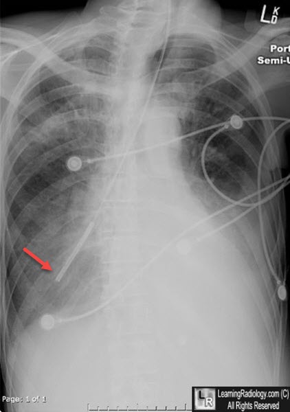
Learningradiology Dobhoff Dobbhoff Tube Malplaced Rll

Abdominal X Ray Revealing The Dobhoff Tube Traversing The Left Main Download Scientific Diagram

Iatrogenic Bronchopleural Fistula From A Dobhoff Tube Radiology Case Radiopaedia Org
Procedure Insertion Of An Oral Nasal Small Bowel Feeding Tube Lhsc

Abdominal X Ray After Fluoroscopic Guided Dobhoff Tube Placement Small Download Scientific Diagram

Icu Chest Radiography Lines Ng Dobhoff Etc Youtube
Dobhoff Nasogastric Tube Tube Radiology Case Radiopaedia Org
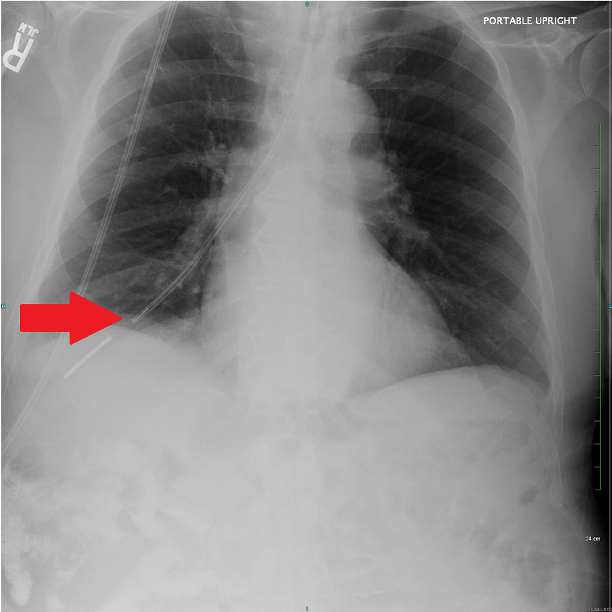
Cureus Hemothorax Following Traumatic Dobhoff Tube Insertion
0 comments
Post a Comment